🖼
FIGURE 2: Visual and retinal features in amyotrophic lateral sclerosis (ALS). The retinal ...
active
paper figure
Created: 2026-04-22T18:14:20
By: paper_figures_pipeline
Quality:
50%
🔗 External
ID: paper-fig-4191f3af-5016-4fac-aa7b-03d1c1
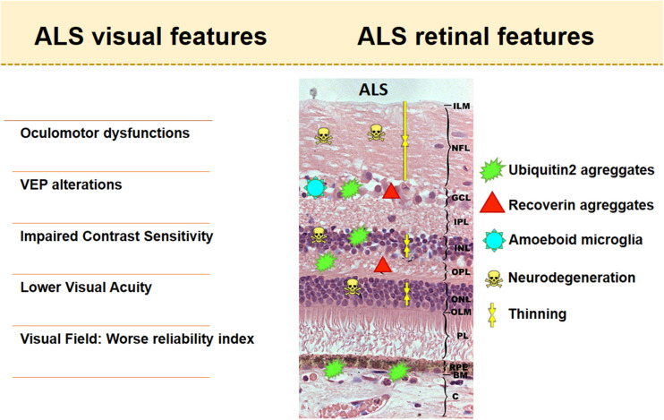
FIGURE 2Figure 2
Visual and retinal features in amyotrophic lateral sclerosis (ALS). The retinal scheme represents the main histological findings found in retinas with ALS.
▸Metadata
| doi | 10.1007/s12011-024-04391-2 |
| pmcid | 7544921 |
| _origin | {'url': 'https://www.ebi.ac.uk/europepmc/articles/PMC7544921/bin/fnins-14-566858-g002.jpg', 'type': 'external', 'tracked_at': '2026-04-23T01:14:20.622207'} |
| caption | Visual and retinal features in amyotrophic lateral sclerosis (ALS). The retinal scheme represents the main histological findings found in retinas with ALS. |
| paper_id | 4191f3af-5016-4fac-aa7b-03d1c17c56fb |
| image_url | https://www.ebi.ac.uk/europepmc/articles/PMC7544921/bin/fnins-14-566858-g002.jpg |
| image_path | |
| description | |
| figure_label | FIGURE 2 |
| figure_number | 2 |
| _schema_version | 1 |
| source_strategy | pmc_api |
| entities_mentioned |
📊 Evidence Profile
Evidence Balance
+0%
Certainty
0%
Debates
0
Incoming
0
Outgoing
0
0 supporting
0 contradicting
0 neutral
Public annotations (0)Annotate on Hypothes.is →
No public annotations yet.